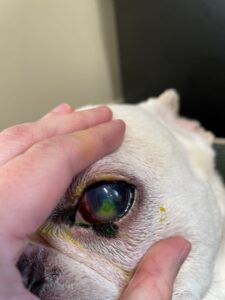
To understand corneal ulcers in dogs, you must first know how the cornea functions.
To understand corneal ulcers in dogs, you must first know how the cornea functions. The cornea is a transparent, shiny component in front of the eyeball. It’s like a clear windowpane. Mount Carmel Animal Hospital is here to help you understand more about corneal ulcers in dogs in this article.
What is It?
The cornea has three layers made of skin cells: epithelium (outermost layer), stroma (cornea’s primary supportive tissue), and Descemet’s membrane (deepest layer). Since these layers of the cornea are transparent, it’s impossible to view them without special stains that color specific cells and emphasize them when the tissue is examined with special lighting.
Corneal erosion or corneal abrasion is the erosion of several layers of the epithelium. A corneal ulcer is a more profound erosion through both the epithelium and the stroma. Furthermore, a descemetocele is a severe progression of an ulcer. This is rare, but if the Descemet’s membrane ruptures, the fluid within the eye, called aqueous humour, can leak out, and the eye itself will collapse.
Several Causes of Corneal Ulcers in Dogs
Although there are multiple causes of corneal ulcers in dogs, the most prevalent reason is trauma. An ulcer might occur due to blunt trauma like a laceration (cat scratch) or a dog rubbing its eye on the carpet. Another common reason is the chemical burn of the cornea. This can happen when an irritating substance like drywall dust or shampoo gets in the eye.
In addition, less prevalent causes of corneal ulcers in dogs include viral infections, bacterial infections, and other diseases. These may start in the eye or form secondary to a condition elsewhere in the body. Examples of other diseases that make a pet more prone to developing corneal ulcers include:
- Keratoconjunctivitis sicca refers to chronic corneal dryness due to limited tear production
- Epithelial dystrophy is the weakening of the cornea
- Endocrine diseases like Cushing’s disease, diabetes mellitus, and hypothyroidism
Diagnosis
A corneal ulcer is painful, and your pet will likely demonstrate that pain through squinting, excessive tear production, scratching at its face, or rubbing its face on things like carpet or furniture. Superficial corneal abrasions are typically invisible without special exams and equipment. However, they are detected with special stains such as fluorescein. After the doctor examines the eye overall, a drop of this stain is placed on the animal’s cornea. The dye will affix to areas of ulceration and become a bright green color. This is made even clearer with a black light to detect any minor scratches or ulcers.
A fluorescein stain test is the most prevalent eye test completed. Also, it might be the only necessary test if the ulcer is acute and superficial. If the ulcer is deep or chronic, samples might be taken for culture and cell study before applying the stain or other medication.
Treatment
The treatment for corneal ulcers in dogs depends on whether a corneal ulcer, corneal abrasion, or descemetocele is present. Mild corneal abrasions typically heal within 5 days with medication to prevent bacterial infections and relieve pain and spasms. However, antibiotic drops are only effective temporarily, so they must be applied often. Your veterinarian will prescribe the appropriate medication and direct you on how often you need to apply it to your pet’s eye.
If a corneal ulcer is present, action must be taken to protect your dog’s eye and promote healing. Besides antibiotic medications, another medication may be added to help promote healing. In more severe cases, it might be necessary to perform a procedure called a corneal debridement to eliminate dead or poorly healing layers of corneal tissue or even to perform a corneal graft. In rare cases, the damage to the eye can be so severe that it requires surgical removal of the eye altogether. Your veterinarian will select the best surgery to maximize healing for your dog.
Risks
The biggest risk with a corneal abrasion is the progression if left untreated. Swift intervention is key to prevent an abrasion from developing into an ulcer or even worse, a descemetocele. Because ulcers are painful, postponing treatment can also result in your pet causing further damage by scratching or rubbing at the eye. A dog will occasionally be sensitive to an ophthalmic antibiotic. If your pet seems to be experiencing pain after using the medication, discontinue it and contact one of the veterinarians at Mt. Carmel Animal Hospital.
HERE AT MOUNT CARMEL ANIMAL HOSPITAL, WE’LL TREAT YOUR PETS LIKE FAMILY!
Mount Carmel Animal Hospital has been serving the Northern Baltimore/Southern York community for over 30 years and is proud to be an independently operated, small animal practice committed to excellence in veterinary medicine and client service. From grooming to wellness services, along with Canine Life Skills Training Courses, and surgical procedures, we have the expertise that will best serve the needs of you and your pet. Contact us at 410-343-0200 and follow us on Facebook!
